



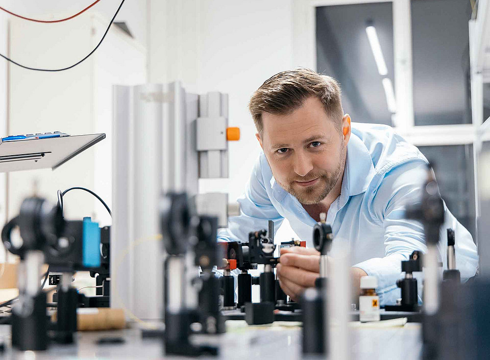
The aim is to identify so-called circulating tumour cells – which detach from tissue and spread throughout the body via the bloodstream – with greater precision. These cells are isolated from blood samples using existing methods. The team is investigating tiny proteins on the surface of these cells. Project leader Jan Enenkel explains: “These proteins are only a few nanometres in size – many thousands of times smaller than a human hair – and can provide important information about the characteristics of the tumour of origin.” Whilst current methods primarily determine which proteins are present and in what quantities, the new method aims to gather more information about the molecular organisation on the cell surface.
Breast and prostate cancer
The focus is, amongst other things, on circulating cells from breast and prostate cancer, which are among the most common forms of cancer. “HER2-positive breast cancer cells are particularly suitable for investigation, as they exhibit a specific surface protein under certain conditions that can be analysed using our technique,” explains Enenkel.
. To achieve this, the physicists are using specially shaped laser light and, at its focal point, a tiny gold nanobead to reveal minute chemical differences on individual tumour cells. A first functional prototype of the analyser is due to be developed over the next 18 months. “In the long term, we want to further develop the technology so that it can provide additional data on cancer cells that is difficult to access using current methods,” says Enenkel, outlining his vision. According to the scientist, the information obtained could one day help to support personalised treatment decisions.